Debunking Your Fears Around Dental Tools and X-rays

One of the scariest things about going to the dentist is the fear of the unknown. Not knowing what tools your dentist will use or why they need you to get X-rays done can feel stressful, but we’re here to debunk your fears about dental tools and X-rays!
X-rays
Radiographic examinations (X-rays) of the mouth and teeth are important to diagnose and manage many different dental problems. Using X-rays, we can identify issues that may not be seen during a routine examination and sometimes even before your symptoms occur.
X-rays are also necessary after trauma to the teeth and jaw as they help your dentist diagnose the full extent of any damage.
Why do I need different types of X-rays?
When it comes to your mouth, you’d be surprised at how many types of X–rays there are! Dentists use different sorts, as each gives us different kinds of information.
We’ve listed some of the most common below.
Bitewings
Bitewings are the most common dental X-ray and are usually used to detect or confirm decay in your teeth and assess the bone level between them. These radiographs also show the crowns of the upper and lower teeth.
Periapical film
These X-rays show the entire tooth, including the root and surrounding bone. They are used for examining the root shape and the area around the root tip, diagnosing bone loss, abscesses and defecting inflammation in the bone due to infections within the root canal of teeth. They are commonly used during root canal treatment, before extractions, and before crowns are prepared.
Panoramic films (OPGs)
OPGs give a 2D view of the entire upper and lower jaws. They give dentists an overall view of your mouth and are particularly useful for identifying the presence of unerupted teeth, wisdom teeth, trauma, and abnormal growths in the jawbone. They have less detail than a periapical or a bitewing and are often required in addition to these. They can also be used to give an overview of developing teeth in children.
Cone Beam Computed Tomography (CBCT)
This type of X-ray produces a three-dimensional image of your teeth, soft tissues and jaw bones in a single scan. It is used when regular dental X-rays are insufficient and is commonly used for locating the origin of pain or pathology, root canal treatment, surgical planning for impacted teeth, dental implant planning, and orthodontics.
What X-ray machine do Mornington Peninsula Dental Clinic use?
At Mornington Peninsula Dental Clinic, we use Intraoral X-ray units that take Periapicals and BWs and an extraoral unit that takes CBCTs and OPGs.
How does a dental professional read an x-ray?
The term “X-ray” refers to the radiation used to create an image – these days read on a computer screen. Tooth decay changes the composition of the hard tooth structures (enamel and dentine) and allows the X-rays to pass through them more. Therefore, a decayed area will appear darker on the images than healthy enamel and dentine. Similarly, inflammation around the root tip of a tooth may destroy bone and appear as a darker area in the image.
Are X-rays safe?
Modern techniques have significantly reduced the risks once associated with X-rays. X-ray equipment must comply with an Australian standard, and dentists must follow strict guidelines. The radiation from an intraoral dental radiograph is less than you receive on any given day from background radiation (radiation from the atmosphere, sun, etc). It is also less than you would receive on an interstate airline flight. There is a known health risk, albeit minimal, associated with all radiographs. However, as in all fields of diagnosis and treatment, it is a case of risk vs benefit.
Dental radiographs and pregnancy
If your dental problems require a radiographic examination, being anxious about any potential harm to you and your baby is understandable. However, the radiation dose and the risks to the unborn child are extremely low. In fact, the failure to treat oral disease may do more harm. According to the National Health and Medical Council, dental radiographic examinations can be done during pregnancy, provided precautions are taken to limit foetal exposure to X-rays by using a lead apron.
If you are thinking of having a baby, having a dental examination before you become pregnant is a good idea. This way, you’ll reduce or avoid needing a dental X-ray examination during your pregnancy.
Tools
What are the most commonly used tools at Mornington Peninsula Dental Clinic?
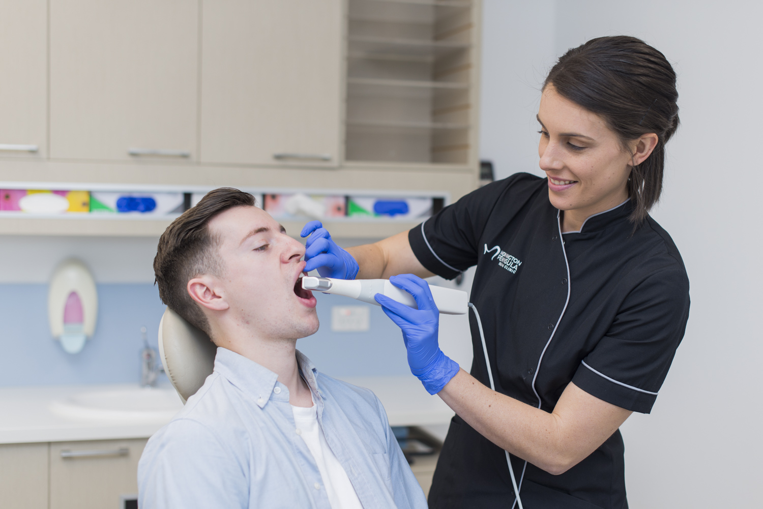
Intraoral scanner
Intraoral scanners are small wand-like devices that digitally reproduce the three-dimensional (3D) structure of both soft and hard tissues (gums and teeth, respectively) inside the mouth. They project a light source onto the surfaces being scanned, and imaging sensors capture the video or images being scanned by the device and produce a 3D model on a computer. Since their induction in 1985, intraoral scanners have continued to develop and have revolutionised dentistry as an alternative to conventional impression methods and have continually gotten faster, more accurate and smaller.
Intraoral scanners have become suitable for a number of clinical applications and include:
- Treatment planning.
- Diagnostics, patient education and health monitoring. E.g. showing areas of concern, orthodontic relapse, teeth movement and the assessment of tooth wear.
- Tooth and implant-supported crowns, veneers, inlays, onlays and bridges.
- Implant planning.
- Orthodontics (braces).
- Removable prosthesis – full or partial dentures.
- Occlusal splints.
- Mouthguards.
One significant benefit of using an Intraoral scanner is the ability to engage patients during an examination. You can show a 3D scan on the computer screen to assist in visualising the current state, potential treatment options and mock-ups and, more importantly, communicate and educate more effectively with the patient.
Numerous studies have shown digital scanning with intraoral scanners to be more favoured by patients in terms of patient-centred outcomes compared to conventional impressions, as they can capture immense detail without using traditional impression techniques, which can sometimes have an unpleasant taste and cause patient gagging.
Check-up and clean tools
Mirror
Typically, a check-up and clean will always include the dentist’s trusty mirror so we can see all areas of the teeth and mouth. We don’t just check the teeth. We check all the structures in the mouth, e.g. tongue, gums, palate, cheeks, and floor of the mouth, to ensure they are healthy. The mirror not only helps us see around corners, but it also helps move tissues and hold them out of the way so we can examine everything properly.
Explorer or “probe”
This is our instrument that has a curved end and comes to a very fine point. It helps us feel around teeth and restorations such as fillings and crowns to make sure they are healthy, and there is no decay, fractures or areas that can catch plaque.
Periodontal probe
This probe with a little ball on the end helps us check your gums. It is run around the gums with very gentle pressure to check for signs of gum disease.
Ultrasonic scaler
This scaler uses ultrasonic vibrations and water to clean the plaque and calculus (tartar) from your teeth. It is a great instrument to get your teeth and gums clean, helping remove stains and preventing and treating gum disease.
Hand scaler
This instrument helps the dentist refine the clean, helping remove any fine bits of plaque and calculus. It has a very fine tip and scrapes along the teeth to remove any remaining plaque and calculus.
Polishing handpiece
This is used along with a brush or rubber cup and fluoride paste after the scaling. It helps remove any biofilm (the first layer of plaque) and any stains and leaves the teeth feeling super smooth and much cleaner.
Camera
The dentists will often take photos of your teeth and mouth to discuss findings with you or to monitor conditions so they can check if they are changing over time.
At Mornington Peninsula Dental Clinic, your comfort is paramount. We know coming to the dentist can bring up a lot of emotions and fears, which is why our team are here to answer any questions you may have about our tools or processes. If you have any questions about your next appointment or to book with one of our team, get in touch today.